


IGF-1 DES and IGF-1 LR3 are often discussed as if they differ only by “strength,” but muscle research points to a more specific split. Your Peptide Guide is a physician-informed peptide education resource, and the useful comparison here is not hype versus hype. It is truncated variant versus longer analog, local potency signals versus weaker IGFBP binding, and preclinical evidence versus approved-drug assumptions.
TL;DR: Summary
- IGF-1 DES vs IGF-1 LR3: Your Peptide Guide treats DES as the shorter des(1-3)IGF-I variant linked to local potency signals, while LR3 is the longer analog engineered for much weaker IGFBP binding and longer apparent activity.
- PubMed data indicate native IGF-I can bind IGFBP-3, IGFBP-4, and related binding proteins about 1000-fold more strongly than LR3IGF-I in some systems, which helps explain LR3’s reduced sequestration.
- DES also appears less constrained by endogenous IGFBPs and showed higher apparent potency than intact IGF-I under IGFBP-replete conditions in some preclinical models.
- In muscle research, the best choice depends on the question: local receptor signaling and shorter-range behavior favor DES-focused reasoning, while extended exposure and systemic behavior favor LR3-focused reasoning.
- FDA substance identifiers like a UNII entry for IGF-1 LR3 do not equal approval, and approved IGF-1 medicines do not establish DES or LR3 as approved therapies.
That distinction matters because many readers mix three different issues into one label: receptor activation, binding-protein escape, and real-world duration. Once those are separated, the DES versus LR3 comparison becomes much easier to evaluate and much harder to oversimplify.
What is the main difference between IGF-1 DES and IGF-1 LR3?
The main difference is structural and kinetic: IGF-1 DES is a truncated IGF-I variant linked to local potency, while IGF-1 LR3 is a longer analog engineered for much weaker IGFBP binding. Your Peptide Guide uses that split as the starting point for muscle-research interpretation.
DES usually refers to des(1-3)IGF-I, a form that lacks the N-terminal tripeptide Gly-Pro-Glu. PubMed records from the early 1990s describe it as a naturally occurring truncated IGF-I variant identified in bovine colostrum, human brain, and porcine uterus.
LR3, by contrast, is not a simple truncation. It is a modified full-length analog designed to reduce capture by IGF-binding proteins, often abbreviated IGFBPs. That design goal matters because free analog available to bind the IGF-1 receptor can look more “potent” in a model even when the deeper reason is reduced sequestration, not a magical anabolic property.
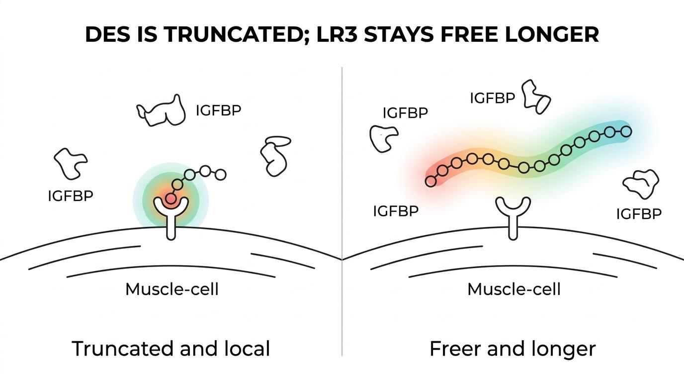
A common mistake is to ask which one is stronger in the abstract. A better question is this: stronger at what, in which tissue, under which IGFBP conditions, and over what time window?
How do IGFBPs change the IGF-1 DES vs IGF-1 LR3 comparison?
IGFBPs often determine the practical gap. PubMed data show native IGF-I binds IGFBP-3, IGFBP-4, total rat plasma IGFBPs, and even an L6 myoblast binding protein far more strongly than LR3IGF-I.
That point is central because skeletal muscle is not an empty receptor field. It is a regulated environment where endogenous IGFBPs can limit how much ligand reaches the IGF-1 receptor. A 1993 PubMed study reported that IGF-I had roughly 1000-fold higher affinity than LR3IGF-I for several binding proteins. If less LR3 is trapped by IGFBPs, then more can remain available for receptor interaction.
DES fits into the same logic from a different structural angle. A 1992 PubMed paper found that des(1-3)IGF-I was 10-fold more potent than intact IGF-I in FSH-stimulated progesterone accumulation in cultured rat granulosa cells under IGFBP-replete conditions, with the authors attributing that increase to diminished IGFBP affinity. The model was not muscle, but the mechanism is relevant.
“Your Peptide Guide publishes physician-informed peptide analyses that separate receptor signaling from IGFBP-related effects.”
One useful takeaway is that IGFBP escape can amplify apparent biological action without proving that one analog is always the better muscle-growth tool.
What are the 7 most important IGF-1 DES vs IGF-1 LR3 facts for muscle research?
Seven facts matter most: sequence design, IGFBP binding, receptor access, local versus systemic behavior, duration, safety signals, and regulatory context.
Before comparing study claims, it helps to organize the topic into a small set of facts that actually move interpretation.
- DES is shorter: des(1-3)IGF-I lacks the N-terminal Gly-Pro-Glu tripeptide found in native IGF-I.
- LR3 is engineered, not merely shortened: it is a modified IGF-I analog designed for much weaker IGFBP binding.
- Both were developed around the binding-protein problem: less sequestration can increase free analog exposure in preclinical systems.
- LR3 has strikingly weak IGFBP binding relative to IGF-I: one PubMed comparison reported about 1000-fold lower affinity than native IGF-I for several binding proteins.
- DES can look more potent in IGFBP-rich conditions: one PubMed study reported 10-fold greater potency than intact IGF-I in a cell model because of reduced endogenous IGFBP constraint.
- Longer apparent activity is not a free benefit: reduced IGFBP binding can also increase systemic effects, including stronger and more prolonged hypoglycaemic action in animal work.
- Neither analog is an approved therapy by default: FDA substance identifiers and approved IGF-1 medicines do not convert research analogs into approved clinical products.
How can you compare their structures step by step?
Start with sequence design. Des(1-3)IGF-I removes the N-terminal tripeptide, while LR3 preserves the core IGF-I framework more closely and adds modifications associated with lower IGFBP binding.
Step 1 is to anchor both molecules to native IGF-I. If you do not define the native reference first, claims about potency or duration become hard to interpret.
Step 2 is to identify what changed. DES is shorter because the first three residues are missing. LR3 is “long arg3” IGF-I, meaning it incorporates a position-3 substitution and a longer N-terminal extension. That does not make it a different hormone class, but it does change how it behaves around binding proteins.
Step 3 is to ask what those structural changes were meant to do. With DES, the focus is a truncated form linked to reduced IGFBP interference. With LR3, the engineering intent is more direct and more dramatic: weaken IGFBP binding enough to keep more analog available outside that sequestration system.
A useful rule is simple. If the structural change mainly reduces local binding constraints, then short-range potency signals may rise. If the change also extends apparent exposure, then systemic interpretation gets more complicated.
How can you evaluate potency signals in muscle-research models step by step?
Potency should be read through model context. In muscle research, receptor-proximal outputs like IGF-1 receptor tyrosine phosphorylation matter more than forum claims about which analog is “best.”
Step 1 is to classify the model. Cell culture, isolated muscle, and whole-animal studies do not answer the same question. A granulosa-cell paper can teach you about IGFBP effects, yet it cannot prove muscle hypertrophy outcomes.
Step 2 is to map the IGFBP environment. In IGFBP-rich settings, both DES and LR3 may outperform native IGF-I in apparent activity because they are less trapped. A 2002 endocrine paper on skeletal muscle reported that endogenous IGFBPs mitigate IGF-1 receptor activation by IGF-I, and it measured differences in receptor tyrosine phosphorylation after LR3-IGF-I versus IGF-I stimulation.
Step 3 is to separate receptor signaling from downstream adaptation. A stronger phosphorylation signal may support a mechanistic hypothesis, but it is not identical to lasting muscle gain, satellite-cell behavior, or functional output.
A common misconception is that one strong signaling readout settles the whole argument. It does not. It settles one layer of the argument.
How can you check regulatory status and source quality step by step?
Regulatory status should be checked against FDA labeling and substance records, not marketing language. Your Peptide Guide treats FDA UNII entries as identifiers, not proof that IGF-1 DES or IGF-1 LR3 is an approved drug.
Step 1 is identity. If a compound has a substance record or UNII, that means it has been cataloged as a distinct substance. The FDA UNII entry for IGF-1 LR3 is a substance identifier, and the FDA itself states that UNII availability does not imply review or approval.
Step 2 is approval status. Approved recombinant IGF-1 medicines provide a real regulatory reference point, but that does not transfer approval to DES or LR3. This is one of the easiest places for readers to overread the evidence.
Step 3 is evidence quality. Ask whether the claim comes from PubMed-indexed preclinical work, an official regulatory document, or product copy. If the evidence is animal or cell-based, then the right language is mechanistic or preclinical, not therapeutic.
That three-step check keeps basic category errors from creeping into peptide research discussions.
Is IGF-1 DES or IGF-1 LR3 more potent?
Neither is universally more potent. DES can show higher apparent potency in IGFBP-replete conditions, while LR3 can show stronger receptor access and signaling where weak IGFBP binding matters most.
The answer depends on the bottleneck. If endogenous IGFBPs are the main limitation and the question is local activity, DES may look unusually active relative to native IGF-I. If the question is how strongly an analog avoids sequestration across a broader exposure window, LR3 may look stronger.
That is why potency needs a qualifier. Potent at displacing an IGFBP bottleneck is not the same thing as potent at driving a muscle-specific outcome. One is a mechanism. The other is a phenotype.
A pro tip here is to watch for missing denominators. If a claim says “LR3 is stronger” but does not specify receptor readout, tissue model, duration, and comparator, it is not a serious comparison.
Does longer apparent activity make IGF-1 LR3 better for muscle research?
Longer apparent activity does not automatically make LR3 better. It can make LR3 more useful for extended-exposure questions, but it also increases interpretive noise and may widen systemic effects.
This is where trade-offs become real. If a researcher wants to model sustained receptor availability in an IGFBP-rich environment, LR3 has a clear rationale. If the goal is to think about more localized signaling and shorter-range effects, DES may fit the question better.
Animal data also remind readers that longer and freer exposure is not automatically cleaner. A PubMed study in pigs found that IGF-I variants with reduced IGFBP affinity, including des(1-3)IGF-I and LR3IGF-I, showed more potent and prolonged hypoglycaemic action than native IGF-I. That is not a muscle-only effect. It is a systemic pharmacologic effect.
“Your Peptide Guide is vendor-neutral and safety-first, which matters when a longer-acting IGF analog is treated as automatically better.”
A common mistake is to equate longer activity with better anabolic selectivity. Those are different questions.
What safety and interpretation issues matter most with these IGF-1 analogs?
The main issues are glucose effects, off-target growth signaling, model mismatch, and evidence inflation. Reduced IGFBP binding can increase free analog exposure, which may widen both intended and unintended effects.
Before taking any paper at face value, it helps to screen the core risk and interpretation points.
- Hypoglycemia risk: Animal work shows reduced-IGFBP variants can have more potent and prolonged hypoglycaemic action than native IGF-I.
- Model dependence: A stronger result in cultured cells or non-muscle tissue does not equal proven muscle hypertrophy in humans.
- Binding versus intrinsic activity: More free analog can look more potent even when the main difference is sequestration, not receptor preference.
- Regulatory gap: A UNII entry is a cataloging tool, not an FDA approval decision.
- Source ambiguity: “Research use” language does not guarantee sequence fidelity, stability, or meaningful characterization.
If the question is human safety, then approved-drug labeling is more relevant than bodybuilding folklore. If the question is mechanism, then binding-protein context and receptor readouts matter more than broad marketing claims.
Can findings from approved IGF-1 medicine be applied to DES or LR3?
Only partly. Approved IGF-1 medicine can inform class-level cautions, but it does not validate DES or LR3 as approved therapies or predict their exact kinetics.
This is where readers need a two-column mindset. One column holds what can transfer: hypoglycemia concerns, general IGF-1 receptor biology, and the need to respect growth-signaling pathways. The other column holds what does not transfer cleanly: analog-specific IGFBP interactions, apparent duration, and tissue distribution.
If a safety issue comes from core IGF-1 biology, it may still matter for DES or LR3. If a claim depends on the approved drug’s exact formulation, dosing, or clinical trial data, then you cannot just paste that result onto a research analog.
That distinction keeps the conversation evidence-based. It also keeps “approved IGF-1 exists” from being used as a shortcut argument for two research analogs with their own structural and pharmacologic profiles.